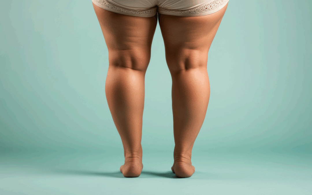
Lipedema is a chronic, progressive and often underdiagnosed disorder of the subcutaneous adipose tissue, with a clear predominance in women. Clinically it is characterized by a symmetrical and disproportionate increase in the circumference of the lower limbs—especially the thighs and calves—and in a significant proportion by involvement of the upper arms, while the palms and soles remain unaffected.
The clinical picture is not the same as simple obesity; pain or tenderness on palpation, a sensation of heaviness, easy bruising and a tendency to develop telangiectasias form the typical symptom complex. The disease becomes apparent or worsens during periods of endocrine change (puberty, pregnancy, menopause), suggesting a hormonal contribution.
Underdiagnosis often leads to repeated diets or intensive exercise without a meaningful effect on the affected limbs, which in turn causes disappointment and emotional distress. Patients also experience stigmatization due to the persistent peripheral disproportionality.
The purpose of this article is to present—within a coherent and evidence-based framework—current knowledge on the pathophysiology, diagnosis and treatment of lipedema, to propose a practical diagnostic algorithm and treatment plan (conservative and surgical), and to delineate the requirements of postoperative rehabilitation and long-term follow‑up.
Definition and Epidemiology
Lipedema is defined as a disorder of fat distribution with a purely peripheral, symmetrical pattern in the lower limbs (and often in the upper arms), accompanied by pain, pressure tenderness and increased ease of bruising.
Prevalence rates vary between studies; double‑digit percentages in the female population are frequently reported, approximately 12%. Symptom onset coincides with major hormonal changes—puberty, pregnancy, menopause—while a positive family history suggests a genetic predisposition.
Underdiagnosis is a public health issue; confusion with obesity and lack of standardized criteria lead to delayed initiation of therapy, during which the disease progresses and symptoms intensify, resulting in a reduction in the patient’s quality of life and often considerable psychological burden.
Early recognition of the entity and its differentiation from lymphedema and other pathologic conditions are prerequisites for effective and safe management.
Etiology and Pathophysiology
Pathogenesis is multifactorial. Genetic factors, hormonal effects (estrogens, progesterone), microvascular instability with increased capillary permeability, subclinical inflammation and partial dysfunction of lymphatic drainage appear to act in combination.
Histopathologically, hypertrophy of adipocytes, heterogeneous lobular architecture, microhemorrhages and fibrosis of the interstitial space are described.
Clinically, this peculiar biology explains the resistance of this peripheral disproportionality to weight loss: even with substantial reduction in BMI, the pathologic tissue in the limbs does not recede accordingly.
Targeted mechanical removal via liposuction acts decongestively and provides permanent management of the pathologic subcutaneous tissue, while conservative methods primarily improve symptom indices (pain, edema, heaviness).


Clinical Presentation and Symptoms
The clinical picture includes symmetrical and disproportionate enlargement of the thighs and calves (often also of the upper arms), pain or tenderness on palpation, a sensation of heaviness and fatigue with standing, easy bruising, frequent telangiectasias and, when present, manifestations of venous insufficiency and varicosities.
In advanced stages, the skin texture becomes heterogeneous with nodular formations and contour deformity—particularly around the knees—resulting in functional limitation of gait.
Functional consequences include difficulty with daily mobility, limitation of sports activities and problems choosing clothing and footwear. The psychosocial burden is significant. Persistent disproportionality and chronic pain undermine self‑esteem and often lead to increasing avoidance of socialization, resulting in the individual’s gradual isolation. Treatment should aim not only at volume reduction but also at functional restoration and pain relief.

Staging
Staging guides therapeutic strategy:
- Stage 1: smooth skin with thickened subcutis, mild symptoms.
- Stage 2: uneven surface with palpable nodules, increased pain and tenderness, more frequent bruising.
- Stage 3: large nodular formations, marked contour deformity, functional limitation.
- Stage 4 (lipo‑lymphedema): coexistence of lymphedema with fibrosis, skin sclerosis and very intense symptomatology that may lead to permanent bed rest.
Accurate staging requires meticulous clinical examination, standardized photographic documentation and, where indicated, imaging studies. Staging influences, the choice between conservative and surgical treatment, which body areas to target, in what sequence, and how many sessions are needed.

Differential Diagnosis
Distinction from obesity and lymphedema is crucial. In obesity there is generalized fat distribution without tenderness of the subcutis. In lymphedema, involvement is often unilateral, includes the foot, and the Stemmer sign is positive.
In lipedema the Stemmer sign is negative; soles and palms are not affected. Venous insufficiency, lipodystrophies and localized lipomatoses are conditions to consider in the differential diagnosis. In complex cases, the final decision is guided by a combination of clinical experience and imaging (ultrasound, MRI).

Diagnostic Algorithm and Imaging
The diagnostic algorithm includes:
- (1) Detailed history (age at onset, hormonal milestones, family predisposition, comorbidities).
- (2) Clinical examination (symmetry of distribution, tenderness on palpation, Stemmer sign, skin texture, signs of venous insufficiency).
- (3) Volumetry, circumferential measurements and standardized photographic documentation.
- (4) Imaging: ultrasound for thickening, subcutaneous hypoechogenicity and increased interstitial fluid; MRI to depict fat signal, fibrosis and possible lymphatic congestion; superficial ICG lymphography in selected cases.
Conservative Management – Principles
Conservative therapy is symptomatic: it reduces edema, pain and the sensation of heaviness.
It is based on four pillars:
- Compression therapy (specialized lipedema stockings, lipedema leggings, sleeves).
- Manual lymphatic drainage.
- Intermittent pneumatic compression (air chambers).
- Lifestyle modification (diet for lipedema, exercise for lipedema).
Correct application and consistency are crucial. Conservative therapy is particularly useful preoperatively (reducing edema, optimizing the skin) and postoperatively (stabilizing the result). In moderate to advanced stages, conservative treatment is insufficient as a sole intervention and should be combined with surgery.
Compression Therapy – Practical Parameters
Compression therapy is a cornerstone. Garments are selected accurately after measuring the affected limbs and choosing the appropriate pressure class. Garments must be applied without folds so as not to impede lymphatic drainage.
Measurements are ideally taken in the morning at specific anatomical landmarks. Duration of use is individualized; in early stages use during physical activity may suffice, whereas in advanced stages extended daily use is required. Garments must be replaced periodically as their elasticity declines.
Patient education on proper application reduces micro‑trauma and discomfort. Regular reassessment ensures that the pressure remains therapeutic as limb volumes change.
Manual Lymphatic Drainage and Pneumatic Compression
Manual lymphatic drainage is performed by specialized therapists and directs lymph toward functioning lymph nodes, reducing edema and pain. It is combined with compression therapy for maximal benefit. Intermittent pneumatic compression devices provide additional decongestion, especially pre‑ and postoperatively.
Parameters (pressure, duration, cycles) are individualized to avoid excessive pressures and discomfort. Integrating both methods within a unified rehabilitation program optimizes outcomes.
Diet and Exercise
Diet does not alter the pathologic tissue of lipedema but supports overall health, reduces fluid retention and thus decreases symptom intensity. A Mediterranean‑style dietary pattern—adequate protein, vegetables, fruits, fish, olive oil, limited salt and sugars—is practically beneficial. Adequate hydration, moderate coffee intake and avoidance of high doses of alcohol help.
Exercise for lipedema should be low‑impact: walking, cycling, swimming, water aerobics, yoga. The aim is to improve microcirculation through mobility, achieving reduction of the heaviness sensation and enhancement of psychological well‑being. Consistency outweighs intensity: low‑intensity, short but frequent sessions are preferable to sporadic intense workouts.
Surgical Management
Liposuction for lipedema is a decongestive and functional procedure. With the goal of permanently removing the pathologic subcutaneous fat, permanent restoration of function and relief from heaviness and pain are achieved.
It is not the same as ‘aesthetic’ liposuction. The operation is performed under general anesthesia for safety reasons, especially when large‑volume removal is anticipated.
Keeping up with scientific advances and applying strict new protocols during the intra‑ and postoperative period, we now safely perform large‑volume liposuction—i.e., removal of more than 5 liters of fat per session—thus achieving faster full‑body treatment with fewer surgeries.
Contraindications and precautions: Contraindications include severe untreated venous insufficiency, poorly controlled diabetes mellitus, active skin infections, or significant hematologic coagulation disorders.
Proper preoperative assessment, evaluation of comorbidities and individualized anesthetic preparation markedly reduce the risk of complications.

Liposuction Techniques and Selection of Areas
Two technologies have prevailed in specialized centers:
- Power Assisted Liposuction (MicroAire PAL), which decreases mechanical resistance and allows precise contouring.
- Water Assisted Liposuction (BodyJet WAL), which facilitates atraumatic tissue separation, although operative time is longer and edema may be greater in some cases.
Area selection follows the principles of unified functional anatomy; knees, calves and ankles are rarely treated in isolation. Typically, the first stage focuses on the posterior compartments of the lower limbs (posterior thighs, calves, ankles, posterior aspect and lateral side of the knee) and, where indicated, the upper arms.
The second stage targets the anterior compartments (anterior–medial thighs, anterior–medial knee). The trochanteric areas (‘saddlebags’) are improved in moderation to preserve gluteal support.
Calves and ankles require high precision for a smooth transition toward the foot and a clear definition of the Achilles tendon. Naturally, the sequence and number of procedures required always depend on an individualized approach based on each patient’s needs.
Safety, Complications and Risk Management
Safety is founded on preoperative assessment, appropriate anesthetic management, meticulous technique and adherence to protocols.
Early complications include seromas, hematomas, transient hypoesthesia, pain and edema, which are managed conservatively.
Late complications may include surface irregularities or persistent tenderness or hypoesthesia; management includes targeted compression, lymphatic drainage or, rarely, corrective intervention. Serious events are rare in organized centers that follow protocols.
Thorough informed consent, realistic expectations, precise preoperative planning and good postoperative care are critical factors for an uncomplicated outcome.
Postoperative Rehabilitation
Immediate application of compression garments reduces edema and stabilizes the surgical planes. Lymphatic drainage sessions in the first 2–6 weeks accelerate fluid resorption and reduce pain and swelling. Intermittent pneumatic compression can be used as an adjunct in selected patients. Pharmacologic support is provided as long as necessary.
Gradual return to activities is medically guided: early gentle mobilization followed by progression to daily activities and low‑impact exercise. Proper application and replacement of garments prevents folds that may cause discomfort and surface irregularities.
Photographic documentation and circumferential measurements enable objective assessment of progress at 1, 3, 6 and 12 months.
Long‑Term Follow‑Up and Prognosis
Reassessments during the first year and annually thereafter evaluate pain, edema, function and quality of life. Maintaining a healthy lifestyle, careful diet, steady low‑impact exercise and, where indicated, continued compression therapy help stabilize results.
Prognosis is favorable when diagnosis is made early and treatment is provided in specialized centers experienced in liposuction for lipedema.
Psychosocial Dimensions
Chronic disproportionality and pain undermine self‑esteem and lead to avoidance of social activities. Destigmatization—clear information that this is an organic disease with distinct pathology—is therapeutic.
Visible improvement after therapeutic liposuction and systematic support reduce psychological burden and enhance adherence to instructions.
Special Situations: Adolescence, Pregnancy, High BMI
In adolescence, timely diagnosis allows conservative measures and education without extreme dieting. During pregnancy, safe compression therapy and gentle exercise with appropriate diet are preferred, avoiding aggressive interventions.
With high BMI, obesity may coexist; weight loss improves some metabolic parameters but does not obviate the need for targeted treatment of the pathologic subcutis. Preoperative evaluation and treatment of venous insufficiency, where present, facilitate the postoperative course.
Quality and Outcome Indicators
Indicative indicators:
- Clinical: reduced pain, reduced sensation of heaviness, increased pain‑free walking distance.
- Objective: reduction in limb circumference and volume, improved skin texture, smoothing of the contour around the knees.
- Patient‑reported: improved quality of life, increased satisfaction with body image, reduced avoidance of social activities.
Systematic collection of these indicators before and after treatment enables evidence‑based evaluation of effectiveness.
Brief Treatment Selection Algorithm
- High suspicion of lipedema: symmetrical peripheral enlargement, tenderness to pressure, negative Stemmer sign → referral to a specialized center for staging.
- Stage 1–mild 2: conservative therapy (compression, lymphatic drainage, exercise, diet) with documentation for 3–6 months and reassessment.
- Moderate 2–3: decongestive liposuction with a clear surgical plan that may include adjunctive procedures where appropriate (e.g., brachioplasty, thigh lift).
- Stage 4 (lipo‑lymphedema): targeted interventions focusing on function, intensification of conservative measures, management of comorbidities.
- In all cases: destigmatization, realistic counseling, recording of outcome indicators, collaboration with multidisciplinary health professionals.
Conclusions
Lipedema is a distinct, chronically progressive disorder of subcutaneous fat with significant functional and psychosocial burden. Diagnosis requires careful clinical examination and targeted imaging where necessary.
Conservative therapy improves symptoms but does not remove the pathologic tissue; surgical decongestion via liposuction is the only evidence‑based intervention for permanent removal with substantial improvement in pain, edema, mobility and quality of life.
Successful outcomes require an experienced surgical team, realistic multi‑stage planning, meticulous postoperative rehabilitation and long‑term follow‑up.
In Greece, access to the specialized clinic of Dr. Daskalakis Dimitrios–Edwardos in Glyfada, Attica enables comprehensive, scientifically documented care for Greek and international patients with lipedema.
References & Documentation
- Van la Parra RFD, Deconinck C, Krug B. Diagnostic imaging in lipedema: a systematic review. Obesity Reviews. 2024;25(1):e13648. https://pubmed.ncbi.nlm.nih.gov/37789512/
- Mortada H, Alaqil S, Al Jabbar I, et al. Safety and Effectiveness of Liposuction Modalities in Managing Lipedema: Systematic Review and Meta-analysis. Archives of Plastic Surgery. 2024;51(5):510–526. https://pubmed.ncbi.nlm.nih.gov/39345998/
- Herbst KL, Zelaya C, Sommerville M, Zimmerman T, McHutchison L. An Advanced Pneumatic Compression Therapy System Improves Leg Volume and Fluid, Adipose Tissue Thickness, Symptoms, and Quality of Life in Women with Lipedema. Life. 2025;15(5):725. https://www.mdpi.com/2075-1729/15/5/725